
Making The Invisible Visible: The Digital Lung Twin

There is one organ we know intimately at Chiesi, yet it remains largely unexplored: the human lung. For Andrea and Georgi, two scientists based at the Paolo Chiesi R&D Campus in Parma, the challenge began with a simple question: could we build a digital representation of the human lung realistic enough to be studied, queried and, one day, used to predict how a therapy might work for each individual patient?
It is an idea that brings together different worlds: industry and academia, pharmaceuticals, mathematics, physics, artificial intelligence and medical imaging. It also reflects the way we approach innovation at Chiesi: not as technology for its own sake, but as a tool to better understand people and develop solutions that respond to their unmet needs.
Can we reconstruct what we cannot see?
Andrea Benassi and Georgi Spasov have dedicated their careers to advancing the science of respiratory drug delivery through computational modelling. Their experience reflects the dynamic nature of innovation, where knowledge is continuously built upon, tested and applied to unlock new possibilities.
Andrea served as Georgi’s scientific advisor during his doctoral research. That collaboration evolved into a continuous research journey, leading them – together with other colleagues and scientific partners – to pursue an even more ambitious goal: not simply modelling part of the lung, but building its digital twin.
Giving the lung a digital identity
A mathematical model can describe a phenomenon. A digital twin aims to do something different: represent a real system in all its complexity, adapting to its unique characteristics and enabling simulations of what could happen under different conditions.
Our project starts with patients’ CT scans. Artificial intelligence identifies the anatomical structures visible in the images, while computational modelling algorithms reconstruct the deeper sections of the bronchial tree that cannot be observed directly. The result is a three-dimensional, patient-specific virtual lung that allows us to simulate the journey of inhaled particles and where they are deposited.
But the ambition extends beyond lung modelling. Our goal is to develop a platform that connects anatomy, breathing patterns, therapies and disease, with the potential to integrate clinical and functional data in the future. This could enable the creation of digital lung models tailored to an individual's physiology and needs, giving each model a distinct identity, as Andrea describes it.

Growing together with the scientific community
A digital twin is not created by a single algorithm. It requires knowledge, collaboration, rigorous validation and ingenuity.
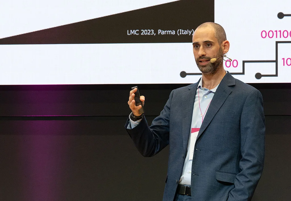
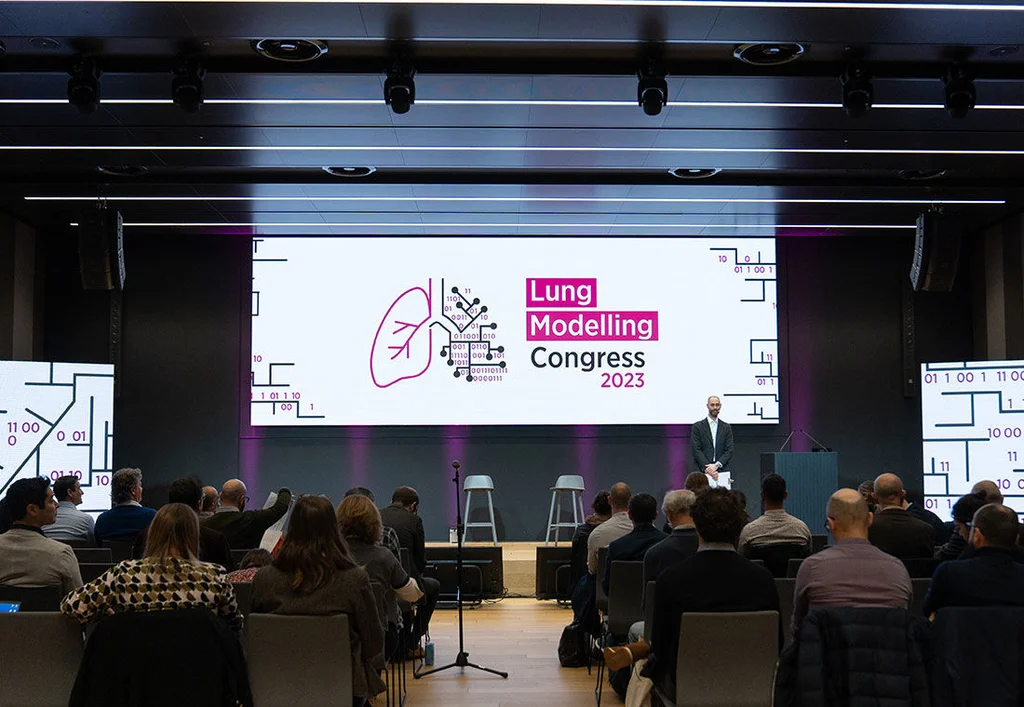
In 2023, Chiesi hosted the first Lung Modeling Congress, bringing together international experts to explore the future of respiratory modelling. Since then, a growing number of scientific publications have documented the project's progress, from modelling aerosol deposition and digitally reconstructing the bronchial tree to building the broader ecosystem needed to make the digital twin increasingly accurate and reliable.
In 2025, our respiratory system digital twin project received a MADE Future Industry Award, recognizing initiatives that bring digital innovation into the industrial world.
From simulation to a new way of doing research
Building the digital twin is only the first step. Before it can be used to support pharmaceutical development, it must be be continuously refined and validated. This requires high-quality data, expertise, close collaboration with clinicians and researchers, and an ongoing dialogue with regulatory authorities.
That complexity is also what makes the project so exciting.
Looking ahead, the digital lung twin could support research aimed at advancing more precise and individualized approaches to respiratory medicine. Achieving that vision will require the same spirit that inspired this program from the start: asking new questions, connecting different areas of expertise, and exploring new possibilities beyond what is currently known.